Photoporation is a physical method of transfection based on the interaction between pulsed laser light and photothermal nanosensitizers. When irradiating the proprietary nanosensitizers with laser light, highly localized light-induced thermal and mechanical forces are generated. When the nanosensitizers are in close contact with the cell membrane, these light-induced forces can form transient pores in the membrane through which external effector molecules can enter the cell. This technology is a next-generation intracellular platform for efficient, flexible and safe delivery of a wide variety of payloads in a broad range of primary and hard-to-transfect cells.
Transfection is a commonly-used method for introducing foreign macromolecules, such as mRNA, proteins and dyes, into cells. Under normal conditions, the cell membrane forms a barrier to prevent this from happening.
Transfection allows foreign payloads to penetrate the cell membrane and enter the cytosol.
There are a variety of techniques used to permeabilize the cell membrane, involving chemicals, electrical pulses or viral vectors, but these are currently expensive, carry risks, and/or are time consuming. See the table below for a comparison between these techniques and photoporation.
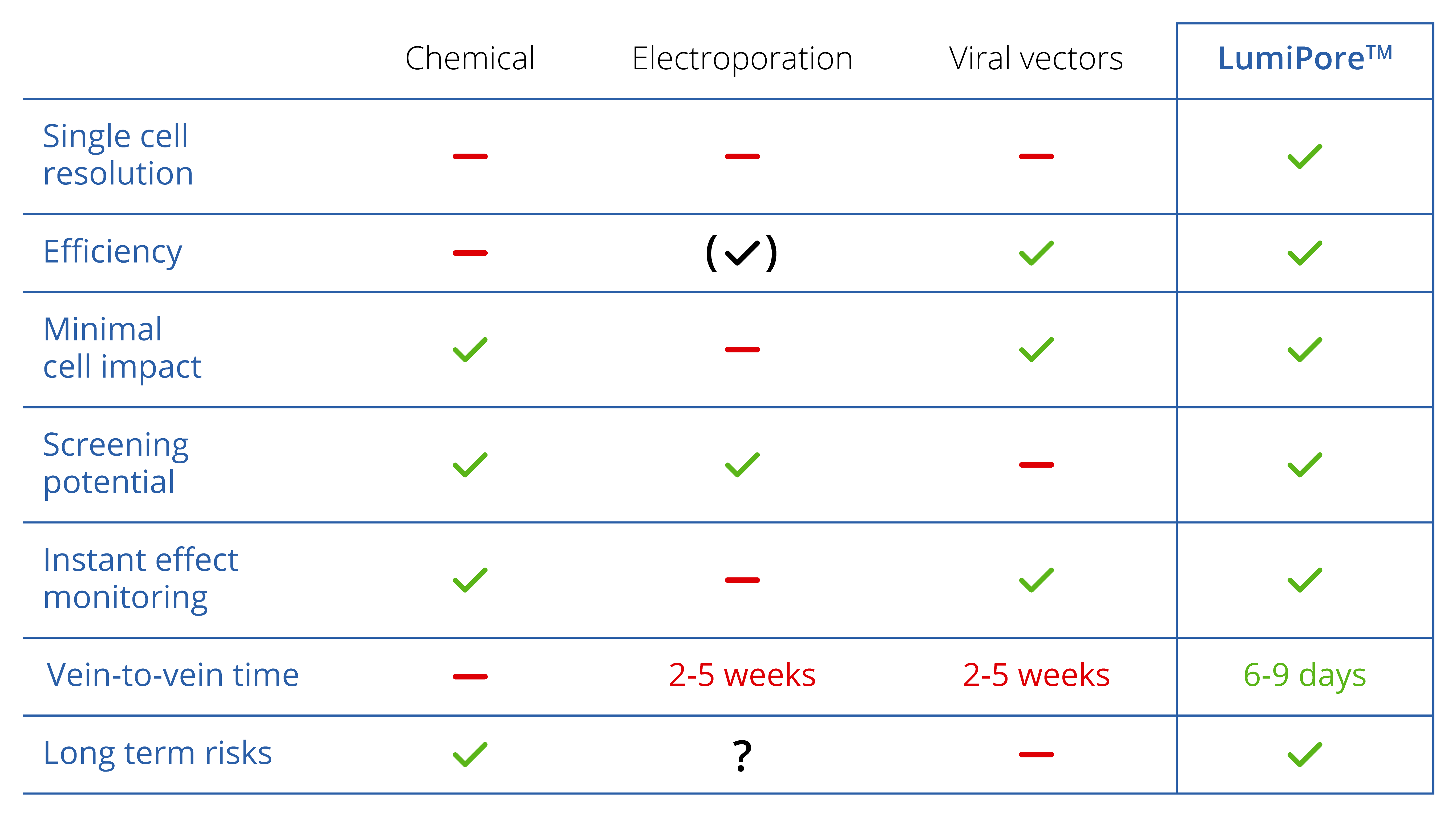
A comparison of transfection capabilities between LumiPoreTM and alternative methods
Learn more about the LumiPore™ photoporation platform
Transfection is commonly used to introduce or alter genes in recombinant cells to study gene function.
The rising popularity of cell and gene therapy is also providing a new use for this technique: adapting human cells for therapeutic use. This obviously requires much more stringent safety standards, since the altered cells will be re-introduced into the human body.
For R&D and clinical applications of transfected cells:
1) Photothermal nanosensitizers and payloads of interest are added to the cell culture medium and incubated with cells.
2) Upon laser irradiation, the cell-associated photothermal nanosensitizers heat up and form nano-vapor bubbles, thus creating pores in the plasma membrane. External macromolecules can enter the cells through these membrane pores.
3) The cells quickly reseal the plasma membrane, resulting in efficiently transfected cells with excellent viability and functionality.
Photoporation uses laser energy to illuminate photothermal nanosensitizers, thus forming pores in the cell membrane and allowing transfection to occur.
Specifically for clinical applications of transfected cells:
In this method, photothermal nanosensitizers are safely embedded into a biocompatible polymer nanofiber structure, shielding them from direct physical contact with cells. The principles and effectiveness of this method are similar to that of our proprietary free-floating nanosensitizers. However, when the cells are collected after laser treatment they are effectively nanosensitizer-free, thus greatly reducing toxicity and regulatory concerns.
Photoporation uses laser energy to heat up photothermal nanosensitizers embedded in nanofibers, thus forming pores in the cell membrane and allowing transfection to occur.
As well as providing an advantageous method of transfecting cells for R&D applications and cell therapies, photoporation has a range of further potential applications such as:
– high throughput delivery of different payloads, including proteins, contrast agents, and genetic material
– light-triggered ablation of cells
– light-triggered delivery or ablation of cells in explanted tissues or even in vivo
Reference: Xiong et al. Adv. Phys. (2016)
Fraire JC, Shaabani E, Sharifiaghdam M, Rombaut M, Hinnekens C, Hua D, Ramon J, Raes L, Bolea-Fernandez E, Brans T, Vanhaecke F, Borghgraef P, Huang C, Sauvage F, Vanhaecke T, De Kock J, Xiong R, De Smedt S, Braeckmans K. Light-Triggered Nanobombs: Nanoscale Biolistics for Efficient Intracellular Delivery of Functional Macromolecules in Mammalian Cells. Nat Comm 2022; 13(1): 2385. (To the article)
Xiong R, Hua D, Van Hoeck J, Berdecka D, Léger L, De Munter S, Fraire JC, Raes L, Harizaj A, Sauvage F, Goetgeluk G, Pille M, Aalders J, Belza J, Van Acker T, Bolea-Fernandez E, Si T, Vanhaecke F, De Vos WH, Vandekerckhove B, van Hengel J, Raemdonck K, Huang C, De Smedt SC, Braeckmans K. Photothermal nanofibres enable safe engineering of therapeutic cells. Nat Nanotechnol. 2021;16: 1281–1291. (To the article)
Harizaj A, Wels M, Raes L, Stremersch S, Goetgeluk G, Brans T, Vandekerckhove B, Sauvage F, De Smedt SC, Lentacker I, Braeckmans K. Photoporation with biodegradable polydopamine nanosensitizers enables safe and efficient delivery of mRNA in human T cells. Adv Funct Mater 2021; 33: Art. 2102472. (To the article)
Raes L, Pille M, Harizaj A, Goetgeluk G, Van Hoeck J, Stremersch S, Fraire JC, Brans T, Gerrit de Jong O, Maas-Bakker R, Mastrobattista E, Vader P, De Smedt SC, Vandekerckhove B, Raemdonck K, Braeckmans K. Cas9 RNP transfection by vapor nanobubble photoporation for ex vivo cell engineering. Mol Ther Nucleic Acids 2021; 25: 696-707. (To the article)
Wayteck L, Xiong R, Braeckmans K, De Smedt SC, Raemdonck K. Comparing photoporation and nucleofection for delivery of small interfering RNA to cytotoxic T cells. J Control Release. 2017 Dec 10;267:154-162. (To the article)
Xiong R, Samal SK, Demeester J, Skirtach AG, De Smedt SC, Braeckmans K. Laser-assisted photoporation: fundamentals, technological advances and applications. Adv Phys-X. 2016;1(4):596-620. (To the article)
Xiong R, Raemdonck K, Peynshaert K, Lentacker I, De Cock I, Demeester J, De Smedt SC, Skirtach AG, Braeckmans K. Comparison of gold nanoparticle mediated photoporation: vapor nanobubbles outperform direct heating for delivering macromolecules in live cells. ACS Nano. 2014 Jun 24;8(6):6288-96. (To the article)
Learn more about LumiPore™
Ottergemsesteenweg Zuid 731 | 9000 Gent | Belgium
Privacy Policy | Terms & Conditions | Cookie policy